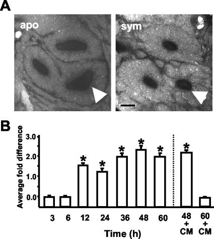
FIG. 6.
Persistent colonization by V. fischeri induced changes in medial duct circumference. (A) Cross-sections through 48-h light organ ducts. The medial duct is indicated with an arrow. Bar, 20 μm. (B) Representative experiment showing the average fold differences, i.e., aposymbiotic dimensions divided by symbiotic dimensions, in the medial duct circumferences. In antibiotic curing experiments (right of the dashed line), cohorts of animals were treated continuously with 20 μg of CM/ml beginning at 12 h. For each condition, n = 15; data are the mean ± the standard error of the mean. A * indicates a significant difference (Student's t test; P < 0.05) between aposymbiotic (apo) and symbiotic (sym) animals.

