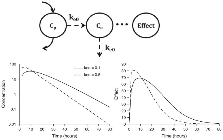
Fig. 4.
Biophase model structure (top panel) and signature profiles for drug concentrations at the biophase (left bottom panel) and pharmacological effects (right bottom panel). Response curves were simulated using Eqs. 1 and 6 driven by drug concentrations following monoexponential disposition: Cp = Coe(−kt). Co was set to 100 units. Parameter values were k = 0.12/h, keo = 0.1 or 0.5/h, Emax = 100 units, and EC50 = 15 units.

