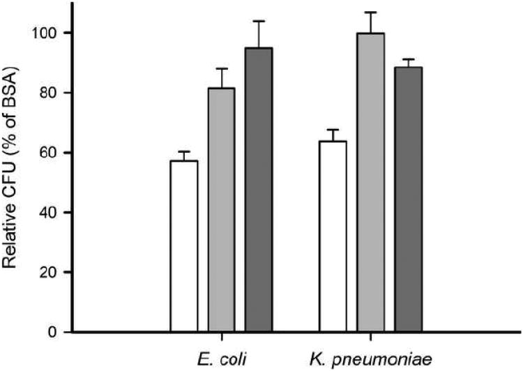
Fig. 9.
Antibacterial activity of apoA-I of E. coli or K. pneumonia. Anti-microbial activity was analyzed by assessing bacterial growth of log-phase gram-negative bacteria in the presence of apoA-I (white bars), ac-apoA-I (light gray bars) and apoA-I Δ190-243 (dark gray bars). Colony counts were relative to BSA (100 %) and were significantly reduced in the presence of apoA-I (57 % and 64 % CFU, respectively). The potency of apoA-I to reduce bacterial growth was significantly decreased after acetylation (82 % and 100 % CFU) or removal of the C-terminal end (95 % and 89 %). E. coli unpaired t-test: WT vs. ac-apoA-I (P = 0.0020, t = 3.849, df = 13); WT vs. apoA-I Δ190-243 (P = 0.0003, t = 4.951, df = 13), ac-apoA-I vs. Δ190-243 (P = 0.2614, t = 1.208, df = 8). K. pneumonia unpaired t-test: WT vs. ac-apoA-I (P = 0.0011, t = 4.285, df = 12), WT vs Δ190-243 (P = 0.0083, t = 3.155, df = 12), ac vs. Δ190-243 (P = 0.2091, t = 1.496, df = 4).

