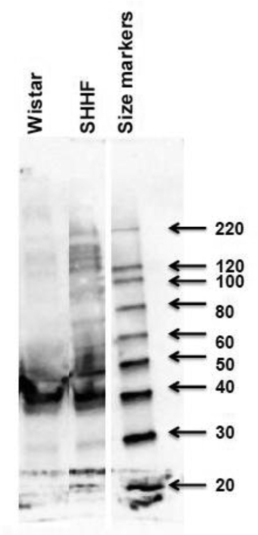
Figure 3. 1D western blot to evaluate increased acetylated proteins in whole cell extracts form W and SHHF rats.
Fifty µg of left ventricular extract is loaded on gel and subjected to western blot using monoclonal anti-acetylated lysine antibody (cell signaling Inc). Molecular weight (kDa) markers are displayed on the right side of the gel.

