
Figure 1.
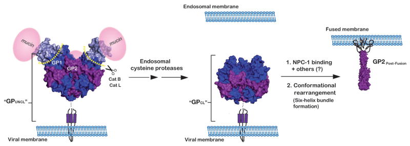
Schematic of the EBOV glycoprotein, GP, throughout its proposed structural transitions during entry into host cells. EBOV GP contains three copies each of a 130 kDa surface subunit, GP1 (blue), and a 24 kDa transmembrane subunit, GP2 (purple). GP1 contains a heavily glycosylated carboxyl-terminal mucin-like domain (represented in pink spheres, “mucin”). GP2 contains a distinct hydrophobic patch at the N-terminus (the fusion peptide) and N-terminal and C-terminal heptad repeat regions (NHRs and CHRs, respectively). After the virus is taken up into the endocytic pathway, endosomal cysteine proteases (cathepsins B and L (Cat B/Cat L) cleave and remove the glycan cap and mucin-like domains of GP1. In response to an unknown trigger, the cleaved GP next undergoes a large conformational change, resulting in the formation of a stable six-helix bundle. Formation of this six-helix bundle is thought to provide the energy required for fusion of the viral and host cell membranes. Here, GP is depicted in its prefusion trimer (PDB ID 3CSY, ref 10), both before and after cathepsin cleavage and in its post-fusion six-helix bundle (GP2Post-Fusion) (PDB ID 1EBO, ref 54). The antigens used for the selection (GPUNCL and GPCL) are soluble forms (i.e. lacking the transmembrane domain) of GP prior to and following proteolysis (respectively). The model for GPCL was generated by modifying the PDB file for GPUNCL (PBD ID 3CSY) to reflect the residues that are removed during cathepsin cleavage (see ref 43).