
Figure 1.
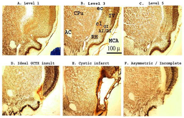
Photomicrographs of NeuN-stained coronal brain sections (2x) through the insular gustatory cortex in Sprague-Dawley rats. As a histological control, the cytoarchitectonic organization of the gustatory and surrounding nuclei was assessed at 5 levels throughout the insular cortex. Sections in A–C, represents the rostral (Level 1), middle (Level 3) and caudal (Level 5) limits of the gustatory cortical zone in an intact SHAM-operated Sprague-Dawley rat, respectively (Levels 2 & 4 not shown). Bilateral ibotenic acid lesions of the gustatory cortex (GCTX) were systematically rated from anterior to posterior in 5 brain sections corresponding to the Levels 1 through 5 (data shown from Levels 1, 3, and 5). D–F. While most ibotenic acid lesions were (D) ideal (limited to the gustatory cortical nuclei), some were (E) overwhelmingly large resulting in cystic infarction, and others (F) were misplaced or asymmetrical (unilateral), thus sparing some gustatory cortical neurons and rated as incomplete. Of the 19 GCTX rats only the behavioral data for 11 GCTX rats, with complete gustatory lesions, were retained for statistical analysis. In total, the data for 8 GCTX rats were discarded due to incomplete lesions (n=3), extensive retrograde damage (see Figure 2 panel D) to the taste thalamus (n=3), or both (n=2). AC: anterior commissure, AI/DI/GI: agranular/ dysgranular/ granular insular cortex, cl: claustrum, Cpu: caudate putamen, ec: external capsule, MCA: (anterior branch of) middle cerebral artery, RH: rhinal horn, IV: cortical layer 4.