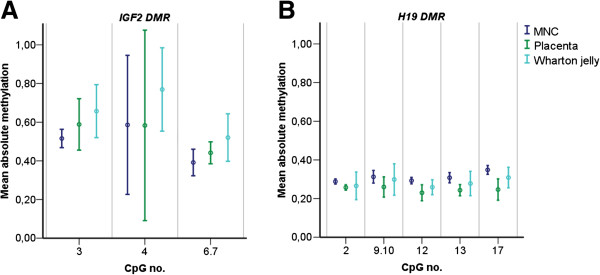
Figure 1.

Mean absolute DNA methylation levels per CpG site for the IGF2 DMR and H19 DMR. Error plots of mean methylation levels (coloured dots) of all individuals ±2 standard deviations (coloured bars) shown for each CpG unit for each of the three tissues separately for (A) IGF2 DMR and (B) H19 DMR. DMR, differentially methylated region; MNC, mononuclear cells.
