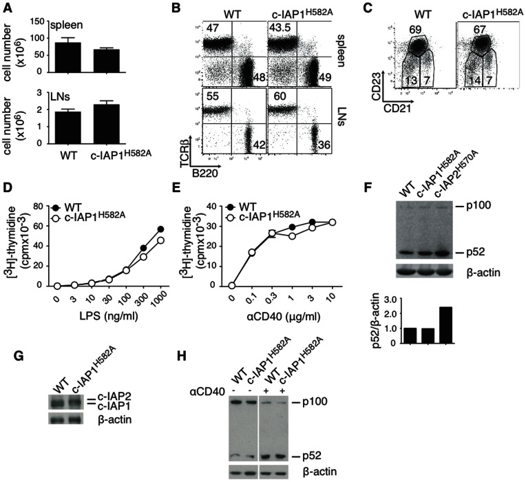
Figure 3. Normal B cell cellularity and function in c-IAP1H582A mice.
(A) Cellularity of spleen and pooled lymph nodes (axial, brachial, superficial cervical and inguinal) from 3 WT and 3 mutant mice. (B) B and T cell distribution was analyzed by flow cytometry. (C) Distribution of marginal zone (CD21hiCD23−), follicular (CD21intCD23hi), and immature (CD21−CD23−) B cells in spleens from WT and c-IAP1H582A mice were analyzed by flow cytometry. (D and E) Purified B cells were cultured in vitro with the indicated concentrations of LPS or anti-CD40 for 48 hr, pulsed with 3H-thymidine, and harvested 18 hr later. (F) Expression of p100 and p52 in purified B cells was detected by immunoblotting. For each sample densitometry of p52 was performed and the results expressed as its ratio to β-actin. (G) c-IAP1/2 expression in freshly purified B cells. (H) Immunoblot of B cells freshly purified or stimulated for 8 hr with 1 µg/ml anti-CD40.

