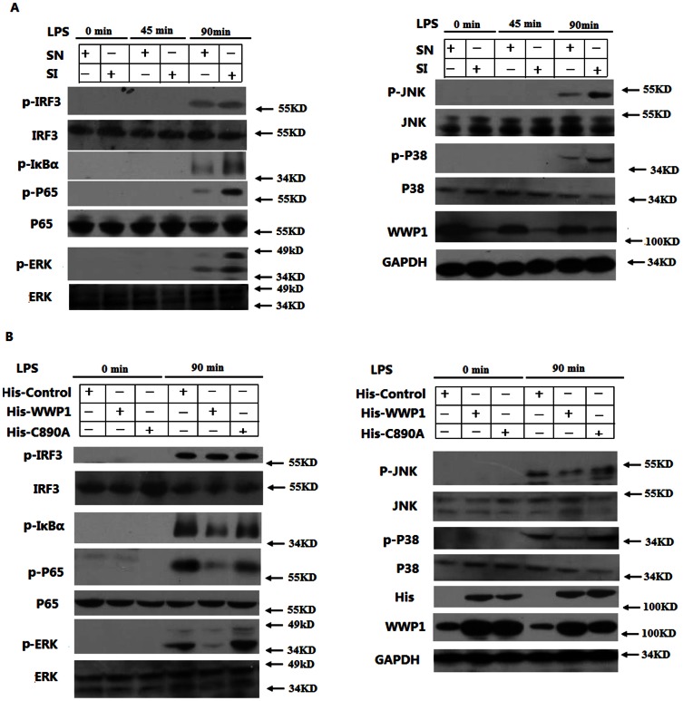
Figure 3. WWP1 inhibits IκB-α, NF-κB, and MAPK, but not IRF3, activation by LPS stimulation.
(A) Knocking down WWP1 enhanced LPS-induced phosphorylation of IκB-α, NF-κB and MAPK, but not of IRF3. Peritoneal macrophages (3×10⁶) were infected with the Lesh NC and Lesh WWP1 virus particles (m.o.i 40) for 48 h, and then, LPS was used (at a final concentration of 400 ng/mL) to stimulate the cells for the indicated times. The lysates were then subsequently analyzed by immunoblots with the indicated antibodies. (B) Over-expression of WWP1 inhibited LPS–induced phosphorylation of IκB-α, NF-κB and MAPK. The RAW264.7 stable cell lines of WWP1 and C890A (4×10⁵) were stimulated by LPS (at a final concentration of 400 ng/mL) for the indicated times. The lysates were then subsequently analyzed by immunoblots with the indicated antibodies. Data are from one experiment representative of three independent experiments with similar results.

