
Figure 2. Fate of injected viable E. coli bacteria in honey bee adult workers.
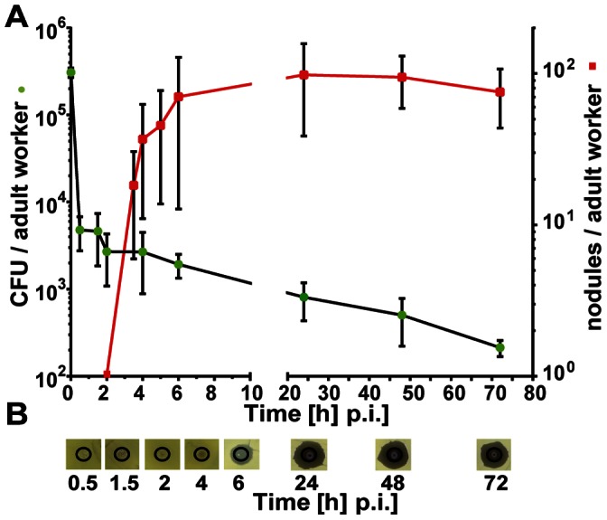
(A) Number of colony-forming units (CFU) recovered from the haemolymph (left y-axis, •) and nodule formation in the haemocoel of infected workers (right y-axis, ▪). Newly emerged worker bees (1d) were challenged with ∼105 E. coli cells and were subsequently divided into two groups: haemolymph was recovered from one batch of individual workers (n = 4–5) at the indicated times (p.i.) and aliquots were spread on nutrient agar plates in order to determine the number of CFUs and the number of melanised nodules per individual worker bee (n = 5–12) was assessed of the second group. The error bars represent the standard deviation. (B) Inhibition-zone assay for the detection of antimicrobial activities in the haemolymph of adult worker bees after infection with E. coli at the indicated times.