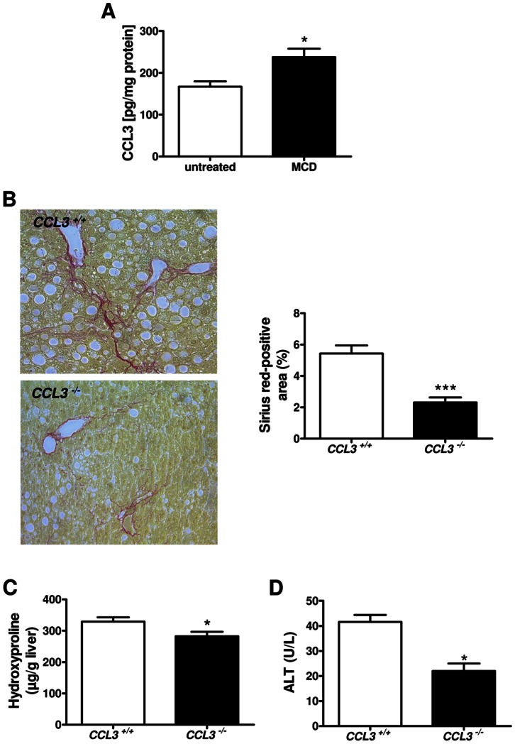
Figure 1. Liver fibrosis in CCL3−/− mice after eight weeks of MCD diet.
CCL3 expression is increased in MCD-fed wild-type mice compared to untreated control (A). Representative Sirius red staining of CCL3−/− and wild-type (CCL3+/+) mice after MCD diet (x100 magnification). Quantitative analysis of the Sirius red-positive area showed a significantly decrease in CCL3−/− mice compared to wild-type mice (B). Hydroxyproline concentration in the liver (C) and serum level of ALT (D) were also significantly decreased in CCL3−/− mice. Data are expressed as means ± SEM of eight mice per group. *P<0.05, ***P<0.001.

