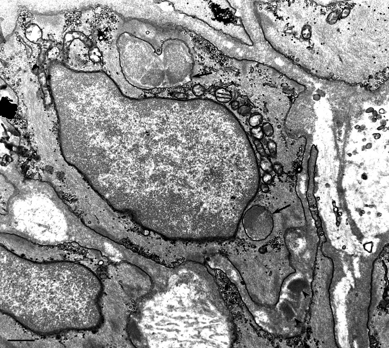
Figure 2. Transmission electron microscopic image of a skin biopsy from a GOM-positive patient (#12).
Two GOM deposits located outside the smooth muscle cell membrane (arrowheads). Due to the plane of section and to the irregular indentations, the cytoplasm of a VSMC contains two GOM pseudoinclusions (arrows). Scale bar = 0.5 µm.

