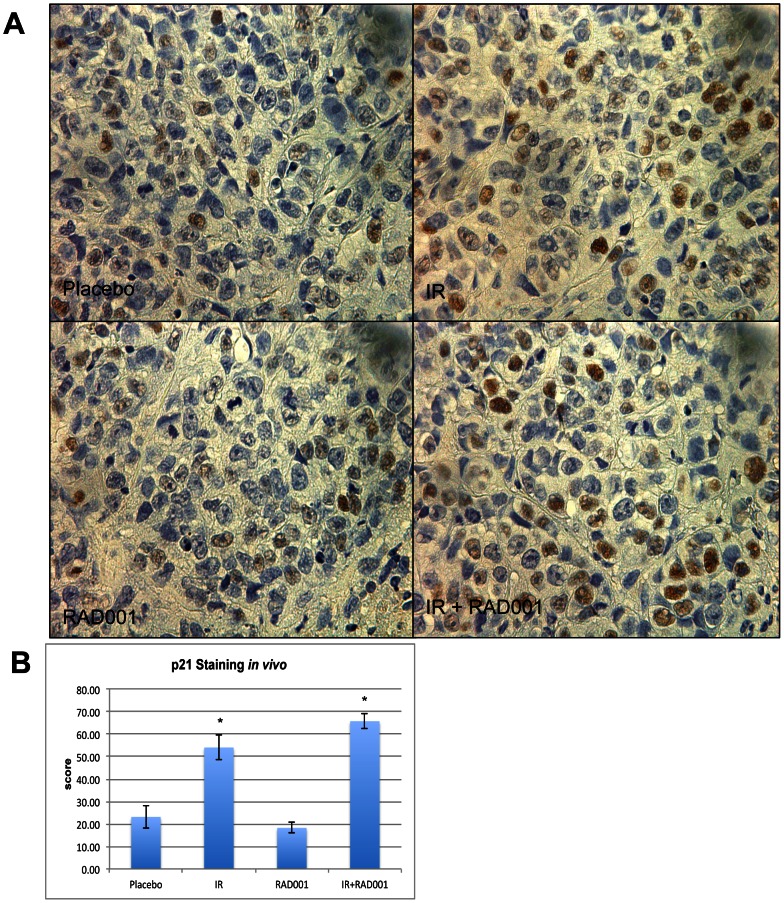
Figure 7. Immunohistochemical p21 levels in mouse xenograft paraffin sections.
(A) Immunohistochemistry was used to detect the levels of p21 in paraffin-embedded mouse xenograft bladder cancer tissues treated with placebo, IR, RAD001 and in combination. (B) Quantification of the immunohistochemistry data revealed a significant increase in p21 expression as observed in tumors treated with ionizing radiation and in combination compared to the placebo and RAD001 treatment.

