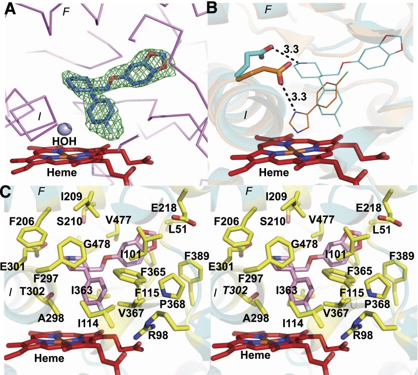
Fig. 3.
(A) An unbiased electron density Fo − Fc omit map obtained before the inclusion of paroxetine in the CYP2B4 structure at 3σ contour level, which clearly shows the presence of paroxetine (blue sticks) above the heme (red sticks). A water molecule (blue sphere) located above heme in the structure is also shown. (B) An overlay of CYP2B4-paroxetine and 4-CPI complexes showing residue E301 in each enzyme, making a hydrogen bond contact at a distance of 3.3 Å from the ligand in the respective structures. (C) Stereo view of CYP2B4 active site residues (yellow sticks) located within 5 Å radius of paroxetine (pink sticks). Heme is shown in red and the protein in cyan.

