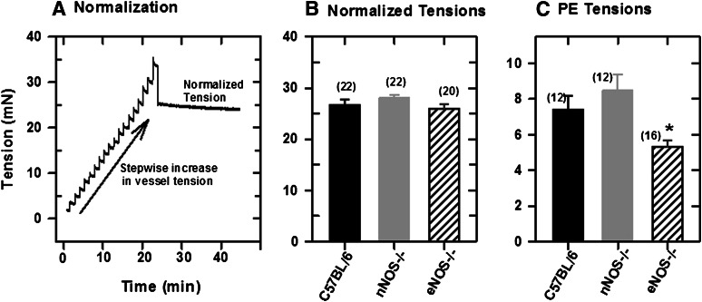
Fig. 2.
Normalization of a mesenteric artery segment mounted in a wire myograph in PSS with 1 mM CaCl2. (A) Normalization: stepwise increase in vessel tension. (B) Normalized tensions in mesenteric artery segments mounted in the myograph chamber. (C) Tensions in normalized vessel segments following applications of 5 μM PE. Values plotted are means (± S.E.M.). Differences in normalized tensions are statistically significant (*P = 0.05; one-way analysis of variance).

