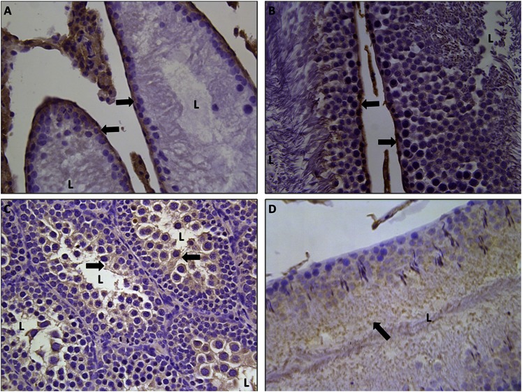
Fig. 7.
Localization of rENT1 and rENT2 in the testis. Immunohistochemical staining for ENT1 (A and B) or ENT2 (C and D) in formalin-fixed paraffin-embedded immature (A and C) or mature (B and D) rat testes is shown at 40× magnification. Arrows indicate positive (brown) staining for proteins. L, lumen of seminiferous tubules.

