
Fig. 5.
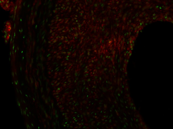
High-power staining of CYP4A1 in balloon-injured (BI) rat carotid artery. The sections were stained with primary antibodies for CYP4A1 and Alexa Fluor 488 secondary antibody. The slide was counterstained with 0.01% trypan blue to quench the endogenous green fluorescence in the tissue. The image shows the positive green staining for the expression of CYP4A1 in infiltrating vascular smooth muscle cells in the neointima.