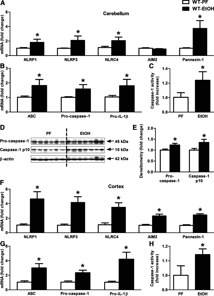
Figure 4. Inflammasome complex is up-regulated and activated in alcohol-fed mice in the cerebellum.
WT mice were fed with an EtOH (n=6) or a control (PF; n=8) diet for 5 weeks. Various inflammasome sensors (NLRP1, NLRP3, NLRC4, AIM2) and Pannexin-1 (A) and the inflammasome adaptor (ASC), the inflammasome effector (procaspase-1), and pro-IL-1β (B) were assessed by real-time PCR from whole cerebellar RNA extracts, normalized to 18S. Inflammasome activity was measured by a caspase-1 colorimetric assay (C) from whole cerebellar lysates. The caspase-1 p10 level (D) was visualized on a Western blot, using β-actin as a loading control, and quantified by densitometry (E). NLRP1, NLRP3, NLRC4, AIM2, and Pannexin-1 (F) and ASC, procaspase-1, and pro-IL-1β (G) were assessed by real-time PCR from whole cortical RNA extracts. Inflammasome activity was measured by a caspase-1 colorimetric assay (H) from whole cortical lysates. Bars represent mean ± sem (*P<0.05, relative to appropriate PF controls by the Kruskal-Wallis nonparametric test).

