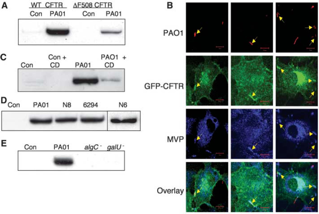
Fig. 1. Recruitment of MVP to lipid rafts of airway epithelial cells by P. aeruginosa.
(A) Lysates of WT and homozygous ΔF508 CFTR human airway epithelial cells (CFT1-LCFSN and CFT1-LC3, respectively) left uninfected (Con) or infected with P. aeruginosa strain PA01-V (PA01) were separated on discontinuous sucrose gradients. Proteins in raft fractions were precipitated and subjected to SDS–polyacrylamide gel electrophoresis (SDS-PAGE) and immunoblot analysis with antibodies directed against human MVP. (B) IB3-1 CF cells (ΔF508/W1282X) transfected with WT-CFTR with an N-terminal GFP tag were infected with CFP-expressing P. aeruginosa, then fixed and stained for MVP. Confocal microscopy identified (arrows) bacteria (red), CFTR (green), and MVP (blue) at the site of bacterial contact with the cell membrane. Scale bar, 10 µm. Overlay shows the merged image of the three individual channels. (C) MVP in rafts of WT-CFTR cells left uninfected (Con) or infected with P. aeruginosa (PA01) in the presence or absence of 5 mM cyclodextrin (CD). (D) MVP in rafts of WT-CFTR cells left uninfected or infected with strain PA01-V or clinical isolates of P. aeruginosa from two CF patients (N6, N8) or from a corneal infection (6294). (E) MVP in rafts of WT-CFTR cells left uninfected or infected with strain PA01-V or LPS mutants of PA01-V (algC− or galU−). The LPS mutants lack the CFTR-binding domain on the bacterial cell surface and do not promote MVP entry into lipid rafts after P. aeruginosa infection.

