Figure 1.
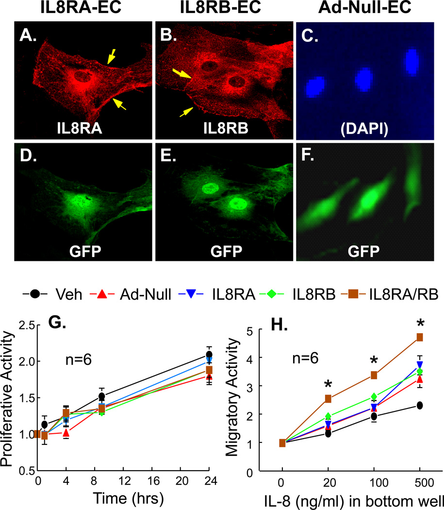
(A-F) Confocal fluorescence micrographs showing over-expression of IL8RA or IL8RB in rat aortic endothelial cells (ECs). ECs were transduced with adenoviral vectors carrying (A, D) IL8RA, (B, E) IL8RB genes, or (C, F) AdNull (EC transduced with the empty adenoviral vector) with a green fluorescent protein (GFP) marker. Arrow heads indicate expression of IL8RA or IL8RB on the EC membrane. The expression of IL8RA, IL8RB, and GFP was controlled by separate CMV promoters. (D, E, F) Immunoflurorescence stain showing expression of GFP in the same ECs. (C) DAPI staining of the nuclei of cells in (F). (G) Proliferative activity (assessed by actual cell counts) of adenoviral transduced ECs over-express IL8RA, IL8RB, or both RA and RB (IL8RA/RB). Vehicle (Veh) control is ECs without adenoviral transduction. Ad-Null control is ECs transduced with empty adenovirus. (H) Migratory activity (assessed by Boyden chamber technique for 12 hrs) of ECs over-express IL8RA, IL8RB or both IL8RA and IL8RB. The bottom wells contain various concentration of IL-8. Results are means±SEM, (n)=wells or chambers. Two way ANOVA was used to analyze the data in 1G and 1H. * p<0.05, IL8-RA/RB vs. other groups in panel H.

