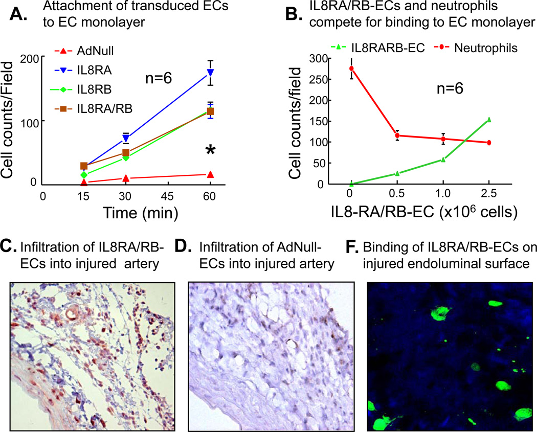
Figure 2.
(A) The attachment rates of IL8RA, IL8RB, and IL8RA/RB ECs to an in vitro endothelial monolayer were faster than that of ECs transduced with empty adenviral vector (AdNull-EC). Endothelial monolayer was stimulated with TNF-α (10 ng/ml) for 16 hrs before AdNull, IL8RA, IL8RB, or IL8RA/RB ECs (with GFP green color) applied to the monolayer. The asterisk indicates that the slope of the lines of IL8RA, IL-RB and IL8RA/RB ECs were steeper (p<0.05) than that of AdNull-ECs. The attachment rates were not statistically different among the IL8RA, IL8RB and IL8RA/RB groups. Results are means±SEM, n=wells/per time point. Two way ANOVA was used to analyze the data in 2A. (B) IL8RA/RB-ECs inhibited binding of neutrophils to in vitro endothelial monolayer in a dose dependent manner. Endothelial monolayer was stimulated with TNF-α (10 ng/ml) for 16 hrs before various concentrations of IL8RA/RB-ECs (GFP color) applied to the monolayer. Neutrophils (labeled with CellTracker CM-Dil, red color) were added 30 min after the application of IL8RA/RB-EC and incubated for additional 1 hr before cell (red and green color) counting in the same well. Results are means±SEM, n=wells/per group. (C) Representative GFP immuno-flurorescence stained micrograph (100X) showing accumulation of GFP-labeled IL8RA/RB-ECs in the injured rat carotid artery (especially in the adventitia and vasa vasorum) 24 hrs after balloon injury of the carotid artery and transfusion of IL8RA/RB-ECs. Transfused ECs were immunostained with GFP primary antibodies. (D) Representative GFP immuno-flurorescence stained micrograph (100X) of GFP-labeled AdNull-ECs in the injured rat carotid artery 24 hrs after injury and cell transfusion. (F) Representative confocal fluorescence micrographs (400X) showing GFP-labeled IL8RA/RB-ECs attached to the endoluminal surface of balloon injured rat carotid artery 30 min after transfusion of IL8RA/RB-ECs. Injured artery was opened longitudinally to expose the endoluminal surface and washed 3 times with PBS before the micrograph was taken.

