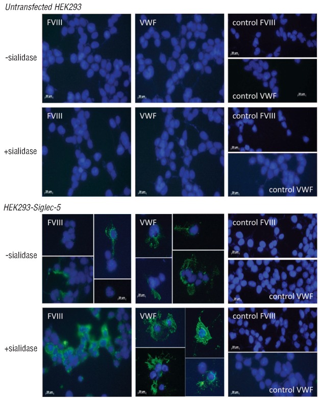
Figure 2.
Binding of FVIII and VWF to Siglec-5 expressing cells. Non-transfected HEK293 cells and HEK293-Siglec-5 cells were grown on glass cover slips and incubated in the absence or presence of sialidase (0.1 U/mL for 1 h at 37°C) prior to incubation with FVIII or VWF (both 10 μg/mL) for 1 h at 4°C. After removing excess of unbound protein, cells were fixed by the addition of methanol. Bound FVIII or VWF was probed using mouse monoclonal antibodies and subsequently detected using AlexaFluor-488 conjugated F(Ab')2 fragments of goat-anti-mouse IgG. Cover slips were then embedded in DAPI-containing mounting medium. DAPI-stained nuclei are presented in blue, while FVIII or VWF are visualized in green. Images were collected using an AxioImager A1 microscope and a Plan-Apochromat 63x/NA 1.4 oil-immersion objective. For control, cells were incubated in the absence of VWF or FVIII.

