
Figure 3.
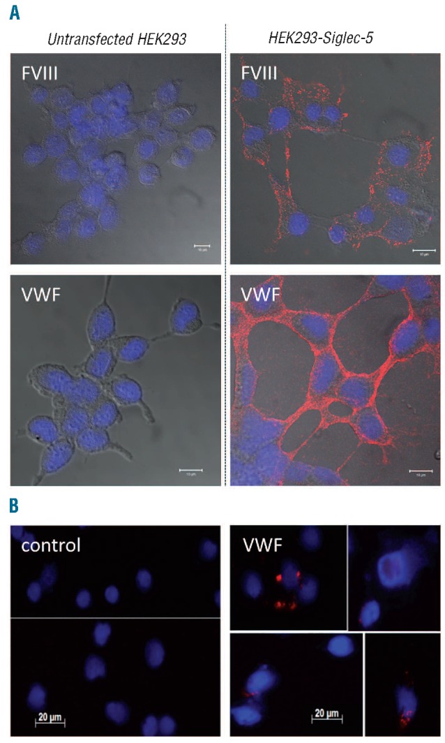
FVIII and VWF co-localize with Siglec-5. (A) Sialidase-treated HEK293-Siglec-5 or non-transfected cells were incubated with FVIII or VWF (10 μg/mL) for 1 h at 4°C. (B) Isolated human monocytes were stimulated with PMA (100 nM) for 1 h incubated in the absence or presence of VWF (10μg/mL) for 1 h at 4°C. (A and B) After removing excess of unbound protein, cells were fixed and incubated with a mixture of mouse monoclonal antibodies against FVIII or VWF in combination with polyclonal goat antibodies against Siglec-5. Bound antibodies were detected via Duolink-PLA analysis by the application of oligonucleotide-coupled secondary antibodies, the sequence of which was complementary. Following amplification, amplified oligonucleotides were highlighted using fluorescent-labeled probes, generating discrete red fluorescent spots for each FVIII/Siglec-5 or VWF/Siglec-5 complex. Cover slips were embedded in DAPI-containing mounting medium to allow blue nucleus staining. Cellular contours are visualized by merging in DIC-images. Confocal-images (A) were obtained with an Axiovert 200M microscope and a Zeiss LSM510-meta confocal system. Regular images (B) were obtained using an AxioImager A1 microscope. In both cases, a Plan-Apochromat 63x/NA 1.4-oil immersion objective was used.