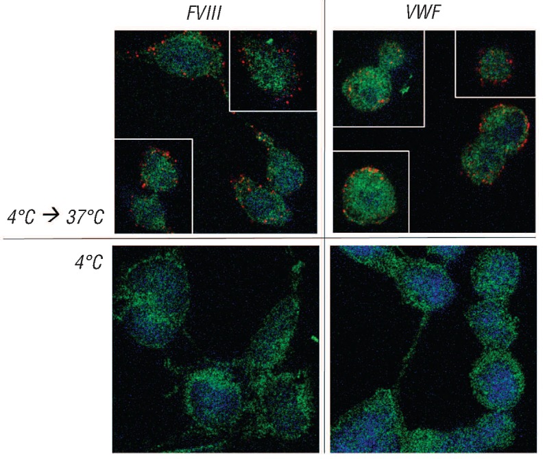
Figure 4.

FVIII and VWF are targeted to early endosomes in Siglec-5 expressing cells. Sialidase-treated HEK293-Siglec-5 cells were incubated with FVIII or VWF (10μg/mL) for 1 h at 4°C. After removing excess of unbound protein, cells were put at 37°C for 30 min in case of FVIII and 15 min in case of VWF, allowing endocytosis of both proteins. Cells were then fixed and incubated with a mixture of antibodies against FVIII or VWF in combination with polyclonal rabbit antibodies against EEA1, a marker for early endosomes. Bound antibodies were detected via Duolink-PLA analysis as described in the legend of Figure 5. Red spots represent FVIII or VWF being within a radius of 40 nM of EEA1. Blue staining represents nuclei. Green staining represents auto-fluorescence of the cells, and is added to visualize the cellular contours.
