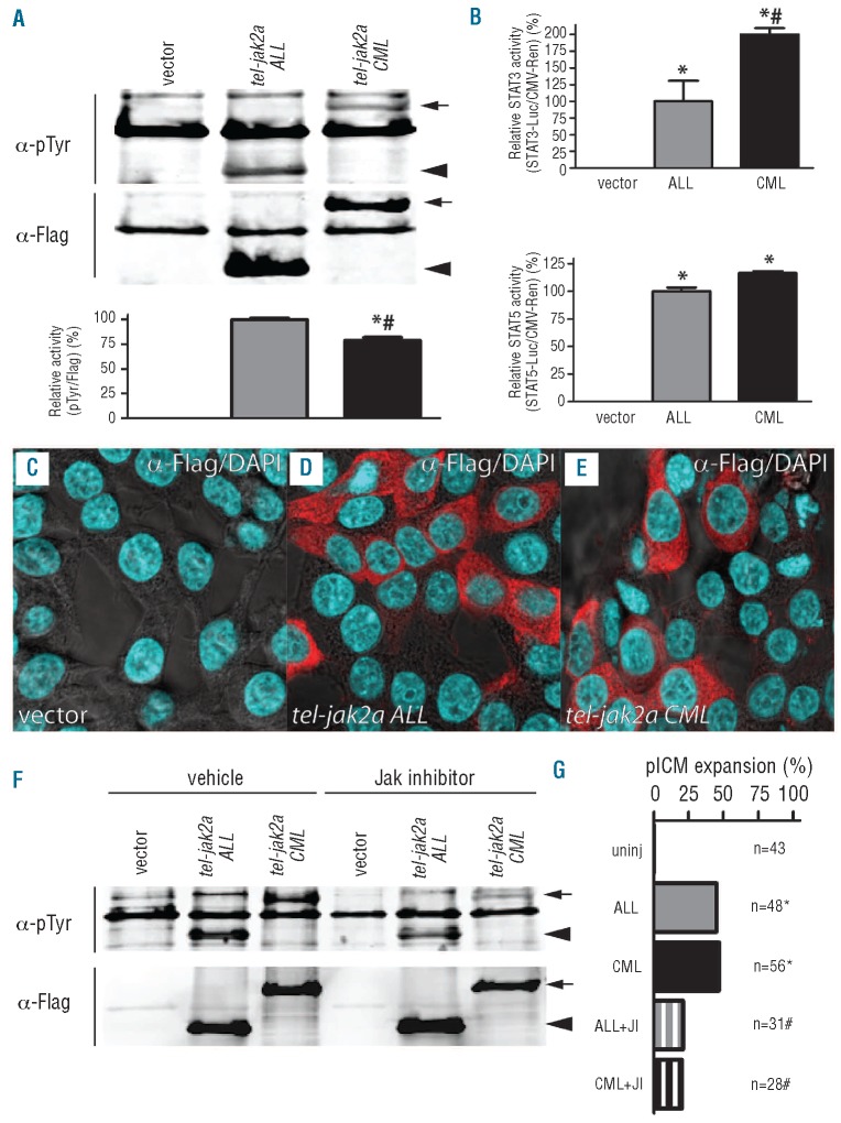
Figure 5.
Functional properties of tel-jak2a fusions. (A) Activity of tel-jak2a fusions. Lysates from HEK293T cells transfected with the indicated constructs were immunoprecipitated with α-Flag and analyzed by western blot with α-phosphotyrosine (α-pTyr) and α-Flag (upper panels), with the relative levels of tyrosine phosphorylation (pTyr/Flag) determined by image analysis, with tel-jak2a ALL set at 100% (lower panel). The tel-jak2a CML protein is indicated with an arrow and the tel-jak2a ALL protein with an arrowhead. (B) Downstream STAT activation by tel-jak2a fusions. HEK293T cells were co-transfected with empty vector, CMV.tel-jak2a ALL and CMV.tel-jak2a CML as indicated, along with STAT-luciferase and CMV.Renilla constructs, with the relative activation of STAT3 (upper panel) or STAT5 (lower panel) reporter determined relative to CMV.tel-jak2a ALL at 100%. (C-E) Localization of tel-jak2a fusions. HEK293T cells were transfected with the indicated constructs and co-stained with α-Flag/Alexa fluor 568nm (red) and DAPI (blue). (F-G) Sensitivity of tel-jak2a fusions to JAK2 inhibitors. HEK293T cells were transfected and analyzed as in panel (A) in the absence or presence of 30 μM AG490 (F). Alternatively, zebrafish were injected with the indicated constructs with or without 30 μM AG490 and scored for posterior intermediate cell mass expansion (G).

