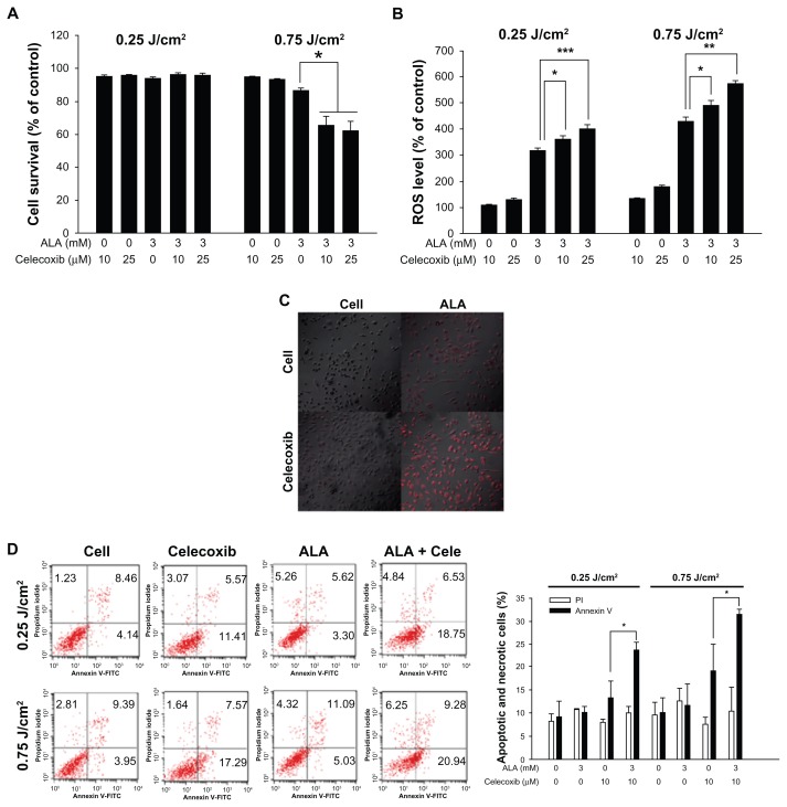
Figure 3.
Cell death and reactive oxygen species (ROS) levels after photodynamic treatment (PDT) with 5-aminolevulinic acid (ALA) and/or celecoxib.
Notes: HuCC-T1 cells were treated with ALA (3 mM) and/or celecoxib (10 or 25 μM) for 4 hours in serum-free medium. (A) After irradiation, the cells were incubated for 24 hours in growth medium containing 10% fetal bovine serum. Cell survival was determined by the MTT assay (error bars represent ± SEM). (B) The ROS level was measured immediately after irradiation. (C) Cells were treated with ALA (3 mM) and/or celecoxib (10 μM). MitoSOX was used to observe ROS generation in mitochondria by confocal microscopy. After irradiation, cells were incubated for 10 minutes at 37°C with MitoSOX (5 μM) and washed with phosphate buffered saline. Cells were fixed with 4% paraformaldehyde and mounted on a slide glass. (D) HuCC-T1 cells were treated with ALA (3 mM) and/or 10 μM celecoxib for 4 hours. Propidium iodide and annexin V were used to detect early apoptosis and necrosis of HuCC-T1 cells. After irradiation, cells were collected immediately and washed with phosphate buffered saline. The collected pellets were stained with propidium iodide and fluorescein isothiocyanate-annexin V and analyzed with a FACScan flow cytometer. Error bars represent ± SEM; * denotes P < 0.05; ** denotes P < 0.01; and *** denotes P < 0.001.
Abbreviations: ALA, 5-aminolevulinic acid; cele, celecoxib.

