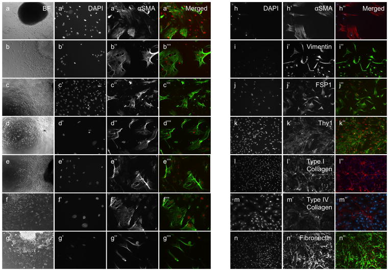
Figure 1.
Derivation of FSMCs from cultured mesothelium. Liver (a–a‴), spleen (b–b‴), kidney (c–c‴), lung (d–d‴), intestine (e–e‴), mesentery (f–f‴), diaphragm (g–g‴). Bright field (a–g), nuclear DAPI staining (a′–g′, h–n). Mesothelium-derived cells express α-SMA (a″–g″, h–h″), Vimentin (i–i″), FSP1 (j–j″), CD90 (k–k″), Collagen type I (l–l″), Collagen type IV (m–m″) and Fibronectin (n–n″). Merged images (a‴-g‴, h″−n″, DAPI is red, αSMA is green except h″, l″, m″). Original magnifications ×20 (a–n).

