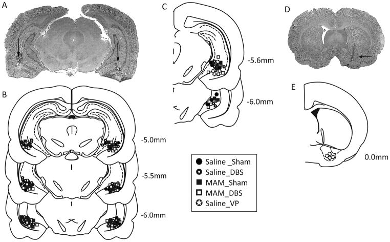
Figure 2.
Histological localization of electrode placements. A representative photomicrograph depicting bilateral electrode locations within the vHipp is depicted in A. The group data for bilateral (B) and unilateral (C) implantations are included schematically. A representative photomicrograph depicting cannula location within the VP is shown in D while the group data are below (E).

