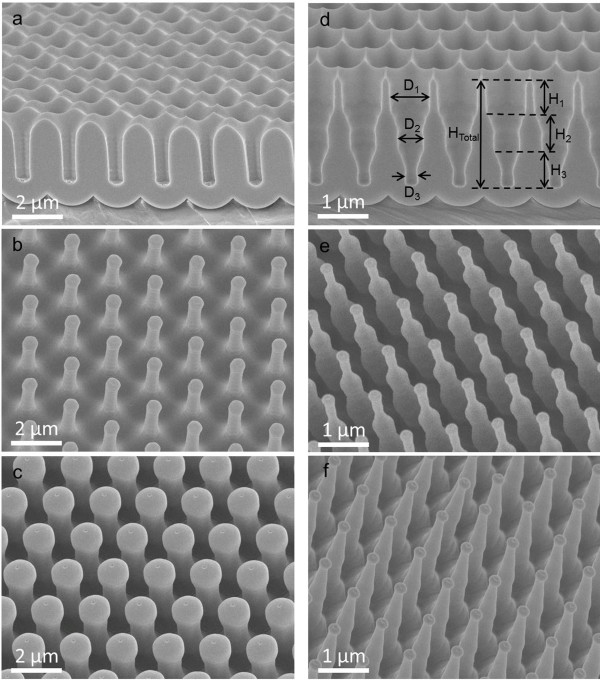
Figure 3.
Cross-sectional-view SEM images of AAM and tilted-view SEM images of PC nanopillar, nanotower, and nanocone arrays. (a) Cross-sectional-view SEM image of 2-μm-pitch AAM with 700-nm pore diameter. The 60°-tilted-angle-view SEM images of (b) PC nanopillar arrays templated from 2-μm-pitch AAM with 700-nm pore diameter, and (c) PC nanopillar arrays templated from 2-μm-pitch AAM with 1.5-μm pore diameter. (d) Cross-sectional-view SEM image of 1-μm-pitch tri-diameter AAM. Tilted-view SEM images of (e) PC nanotowers and (f) PC nanocones.

