
Figure 1.
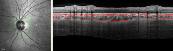
Images from 360-degree 3.4 mm diameter peripapillary circle scans. Examples of images describing choroidal thickness and demonstrating manual delineation of the choroidal vasculature lying between the outer border of the retinal pigment epithelium (RPE) and the inner surface of the sclera.