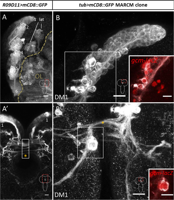
Figure 1.

Type II neuroblast lineages labeled by R09D11-Gal4 and mosaic analysis with a repressible cell marker (MARCM) in the late larval brain. (A,A’)R09D11-Gal4 driven labeling of eight dorsomedial (DM) lineages and their commissural fascicles. DM1 to DM6 indicated with numbers; lat, lateral type II lineages. Z-projection of multiple adjacent optical sections. (A) shows one hemisphere, (A’) shows the midline regions of the larval brain with the commissure indicated by orange asterisk in (A’) and part of the commissural region overexposed to show fiber tracts (white box in A’). (B,B’)tubulin-Gal4 driven MARCM-based labeling of DM1 lineage shows neurons and glial cells in a single neuroblast clone. Axonal fascicles from the DM1 clone cross the commissure (asterisk in (B’)). Z-projection of multiple optical sections. The slightly larger cells at the base of the lineage are glial cells expressing gcm-lacZ (insets in (B,B’); red, gcm-lacZ). Scale bars, 10 μm.
