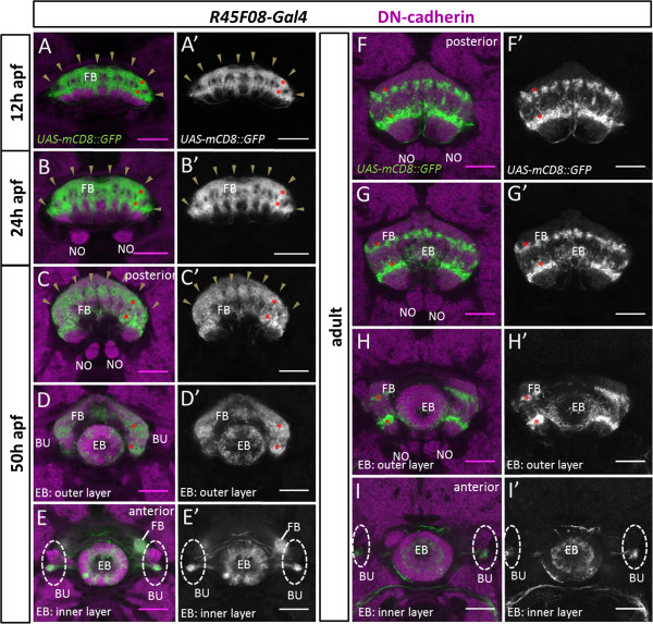
Figure 8.
Innervation of modular subdomains in the developing central complex neuropile by R45F08-Gal4-labeled neurons. Spatiotemporal analysis of the modular innervation pattern of R45F08-Gal4 expressing cells in the developing central complex during metamorphosis and in the adult. R45F08-Gal4 labeling (green), DN-cadherin labeling of neuropile (magenta); all panels show single confocal sections. (A,A’) At 12 h after puparium formation (apf) R45F08-Gal4-labeled innervation of the developing fan-shaped body manifests eight columnar domains (arrowheads) arranged in two closely apposed horizontal layers. (B,B’) At 24 h apf R45F08-Gal4-labeled innervation of the developing fan-shaped body has grown but continues to show eight columnar domains (arrowheads) arranged in two closely apposed horizontal layers. (C-E’) At 50 h apf R45F08-Gal4-labeled innervation of the fan-shaped body has expanded further but is still seen in eight columnar domains (arrowheads) arranged in two horizontal layers. Labeled innervation is also seen in the inner and outer layer of the ellipsoid body, and innervation is seen in the ventral part of the bulbs. (C,C’,D,D’,E,E’) are taken at different focal planes along the A/P axis in the developing central complex. (F-I’) In the adult R45F08-Gal4-labeled innervation of the mature fan-shaped body is seen in two distinct layers that are clearly separated by unlabeled neuropile. Whereas in the ventral layer the eight columnar domains seem to have fused, in the dorsal layer the columns appear to be further subdivided into a 16-fold modular organization. Labeled innervation remains in the inner and outer layer of the ellipsoid body and the ventral part of the bulbs. (F,F’,G,G’,H,H’,I,I’) are taken at different focal planes along the A/P axis in the central complex. In (A-D’,F-H’) asterisks indicate one labeled columnar domain in two layers of the fan-shaped body. BU, bulbs; EB, ellipsoid body; FB, fan-shaped body; NO, noduli. Neuroanatomical nomenclature according to [3,40]. Scale bars, 25 μm.

