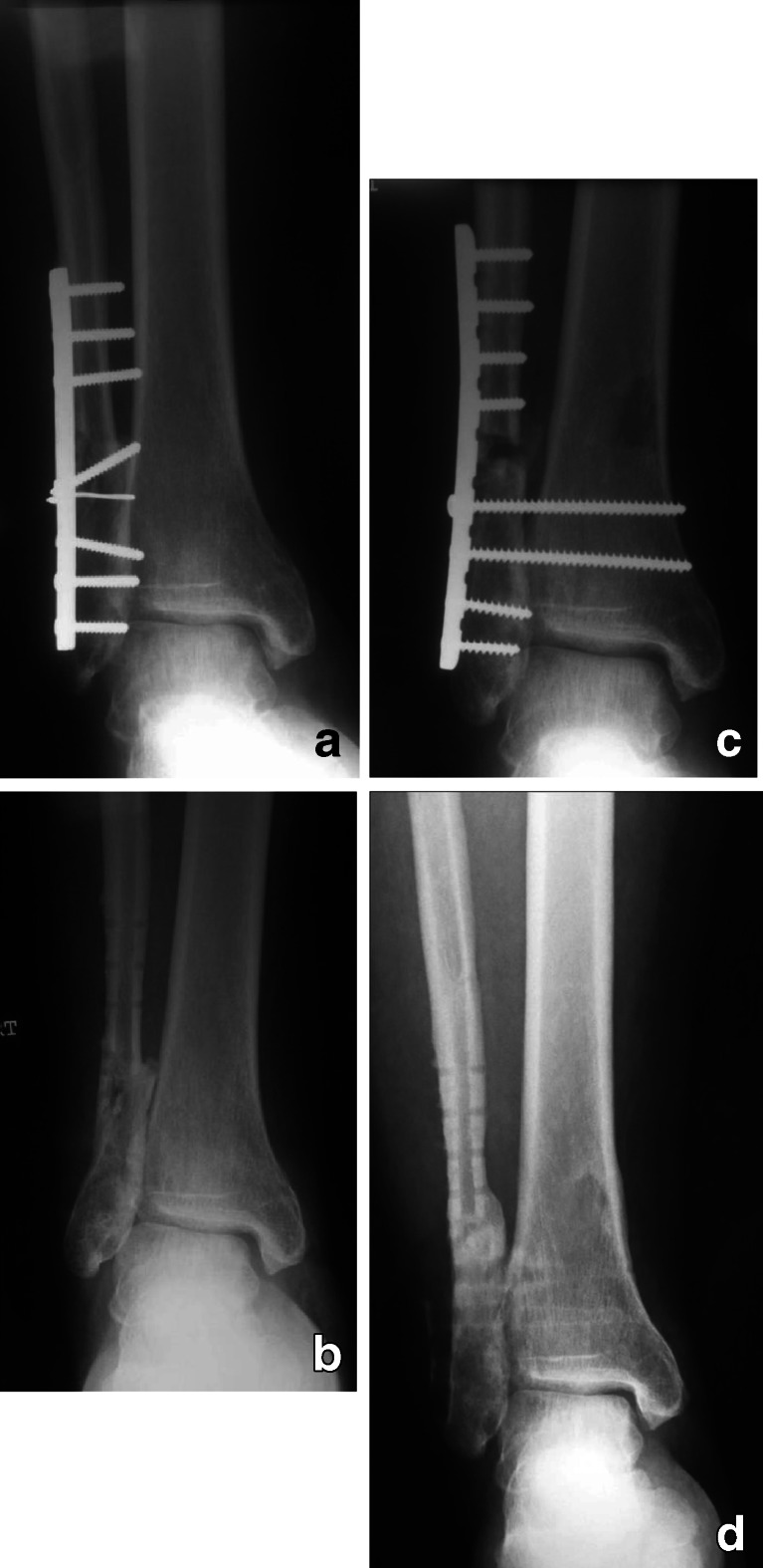
Fig. 1.
(a to d) a Radiographs of an ankle fracture treated by open reduction and internal fixation. The lateral malleolus was fixed in shortening and under-reduced. b Radiographs after implant removal show the lateral malleolus is united in external rotation and shortening, the medial clear space is widened and radiological evidence of osteoarthritis K-L grade 2. c Post-operative radiographs after reconstructive surgery. The fibular length and alignment are restored, the medial clear space is reduced to normal, and the resultant osteotomy gap is filled with a graft harvested from the ipsilateral distal tibial metaphysis. d Follow up radiographs after complete healing and implant removal

