
Fig. 4.
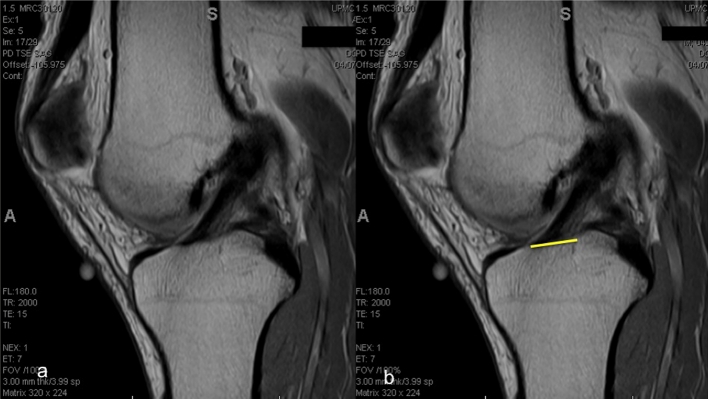
a Sagittal MRI cut best showing the ACL attachment to the tibia; b the most anterior and most posterior fibres attaching the tibia are connected by a line that represents the insertion site size
Official websites use .gov
A
.gov website belongs to an official
government organization in the United States.
Secure .gov websites use HTTPS
A lock (
) or https:// means you've safely
connected to the .gov website. Share sensitive
information only on official, secure websites.
a Sagittal MRI cut best showing the ACL attachment to the tibia; b the most anterior and most posterior fibres attaching the tibia are connected by a line that represents the insertion site size