
Fig. 5.
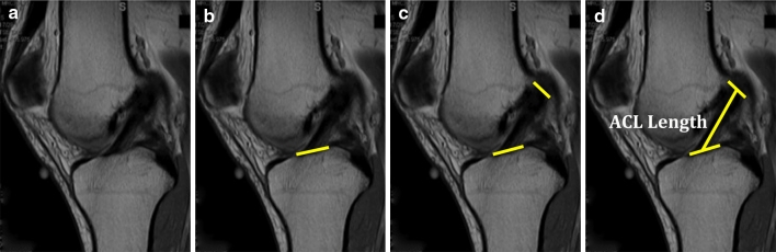
a MRI sagittal cut best showing tibial and femoral insertion sites is chosen; b tibial insertion site is highlighted; c femoral inserion site is highlighted; d the distance between the mid-portion of the tibial and femoral insertion sites are connected and measured to know ACL length