Fig. 5.
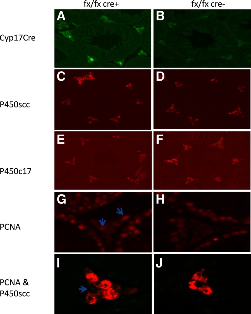
Immunohistochemistry for P450scc, StAR, P450c17α, and PCNA in Leydig cells. A and B, 17-Hydroxylase-cre-IRES-GFP is expressed in Leydig cells of ALK3fx/fxCyp17cre+ mice (A) but not control mice (B); C–F, although P450scc expression is similar (C and D), P450c17α staining intensity is lower in ALK3fx/fxCyp17cre+ (E) than control (F) mice.; G–J, PCNA expression (arrows) is significantly higher in ALK3fx/fxCyp17cre+ (G and I) than control (H and J) mice.

