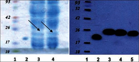
Figure 3.

(A) SDS-PAGE analysis of bacterial lysate extract. Lane 1, molecular weight marker; Lane 2, standard GH, Lane 3,4 bacterial lysate extract before purification; (B) Western blot analysis of bacterial lysate extract. Lane 1, molecular weight marker; Lane 2, standard GH, Lane 3,4 bacterial lysate extract before purification; Lane 5, bacterial extract after purification by His·Bind Quick 300 Cartridg column. The protein ladder has been highlited using pen marker for better visulalization.
