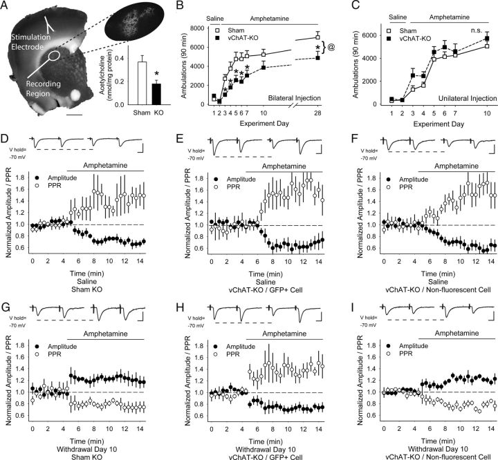
Figure 10.
Effects of partial acetylcholine depletion in the dorsal striatum. A, Coronal corticostriatal slice showing the areas of stimulation and recording with histology and the typical location of AAV1-Cre-GFP. Inset: Tissue acetylcholine content in the dorsal striatum of sham-KO and vChAT-KO injected with AAV1-Cre-GFP. *p < 0.05, Student's t test. B, Locomotor ambulations in sham-KO and vChAT-KO mice after bilateral injections with AAV1-Cre-GFP. *p < 0.05, Student's t test; @p < 0.05, 2-way ANOVA. C, Locomotor ambulations in sham-KO and vChAT-KO mice after unilateral injections with AAV1-Cre-GFP. D, Representative traces (top) show the average responses of cortically evoked paired pulses before (left) and 5 min after bath application of amphetamine (right). Amphetamine reduced the eEPSC amplitude and increased the PPR in MSNs from saline-exposed, bilaterally injected sham-KO mice. E, F, In saline-exposed, bilaterally injected vChAT-KO mice, amphetamine reduced the eEPSC amplitude and increased the PPR in both GFP+ MSNs (E) and in nonfluorescent MSNs (F). G, Amphetamine increased the eEPSC amplitude and decreased the PPR in MSNs from amphetamine-treated, bilaterally injected sham-KO mice. H, Amphetamine reduced the eEPSC amplitude and increased the PPR in GFP+ MSNs from amphetamine-treated, bilaterally injected vChAT-KO mice. I, Amphetamine increased the eEPSC amplitude and reduced the PPR in nonfluorescent MSNs from amphetamine-treated, bilaterally injected vChAT-KO mice. Scale bars in A, 1 mm; D–I, 100 pA, 5 ms.

