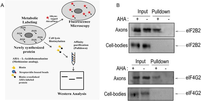
Figure 3.
mRNAs encoding eIF2B2 and eIF4G2 are translated in the distal axons of SCG neurons. A, A schematic diagram showing the metabolic labeling strategy used to detect newly synthesized axonal proteins. AHA, an analog of l-methionine, was added to axonal compartments, biosynthetically incorporated into newly synthesized proteins, and detected by a copper (I)–catalyzed cycloaddition (Click-iT) reaction to either an alkyne-derivatized fluorophore or biotin-alkyne followed by streptavidin affinity absorption and Western analysis. Local synthesis of eIF2B2 and eIF4G2 proteins was assessed by metabolic labeling of axonally synthesized proteins with AHA for 6 h, followed by biotinylation and affinity purification by streptavidin beads. As control, soma lysates from center compartments whose axonal compartments were labeled with AHA were also subjected to biotinylation and affinity purification by streptavidin beads. B, A Western analysis was performed on the affinity-purified fraction to detect the presence of eIF2B2 and eIF4G2 using monoclonal antibodies. There is a lack of AHA-labeled (pulldown fraction) eIF2B2 and eIF4G2 in the lysates from the cell bodies. Ten percent of the total input and 50% of the pulldown were loaded in each lane of the SDS gel for the Western analysis.

