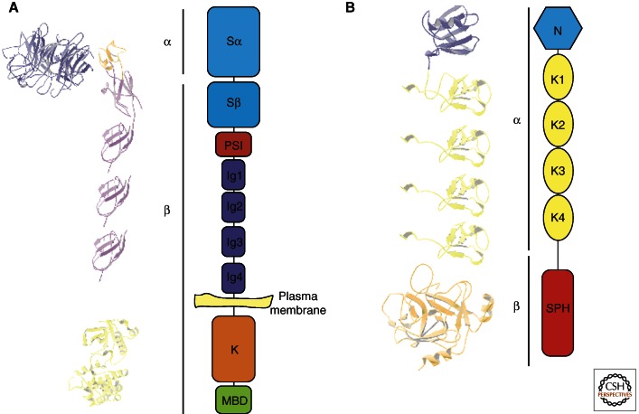
Figure 1.
Structural characteristics of MET and HGF/SF. (A) Structure of the MET receptor (α and β refer to subunits present after proteolytic cleavage). MET is expressed at the plasma membrane: The extracellular portion consists of the sema domain, a PSI domain, and four immunoglobulin-like (Ig-like) repeated domains; the intracellular region contains the tyrosine kinase domain and the multifunctional binding domain. The three-dimensional models (left) were generated using the following coordinates from the Protein Data Bank: 1SHY (sema domain), 2UZY (PSI and Ig1, and 2), and 1R1W (kinase domain). The Ig3 and Ig4 domains are modeled as copies of the Ig2 domain. (B) Functional domains of HGF/SF. HGF/SF contains an amino-terminal domain (N), four tandem repeats of kringle domains (K1–K4), and a serine protease homology domain (SPH). The three-dimensional models (left) were generated using the following coordinates from the Protein Data Bank: 1NK1 (N and K1) and 1SHY (HGF/SF β chain). K2–4 are represented as copies of K1.

