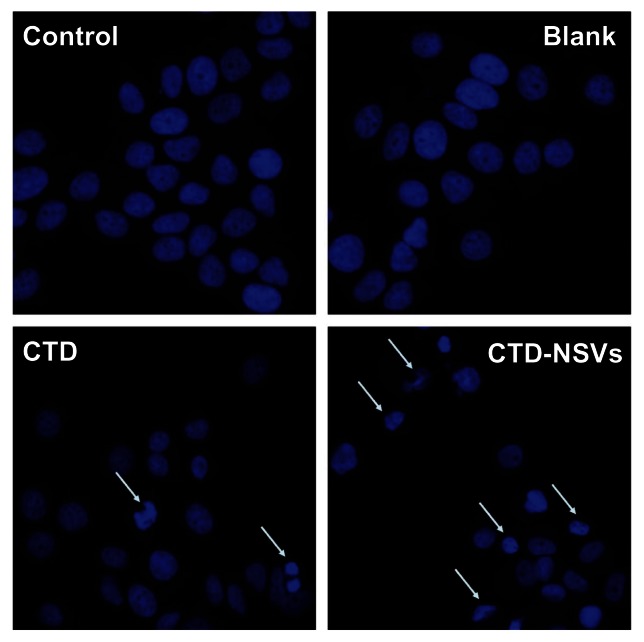
Figure 4.
Hoechst 33342 fluorescent staining to detect apoptotic morphology in breast cancer cells.
Notes: MCF-7 cells were treated with blank non-ionic surfactant vesicles (NSVs), free cantharidin (CTD) (20 μM), and CTD-NSVs (20 μM) for 48 hours. Cells were observed in three experiments using a fluorescent microscope (Olympus, Tokyo, Japan).
Abbreviation: CTD-NSVs, CTD-loaded non-ionic surfactant vesicles.

