Figure 1.

Example of evaluated CT:
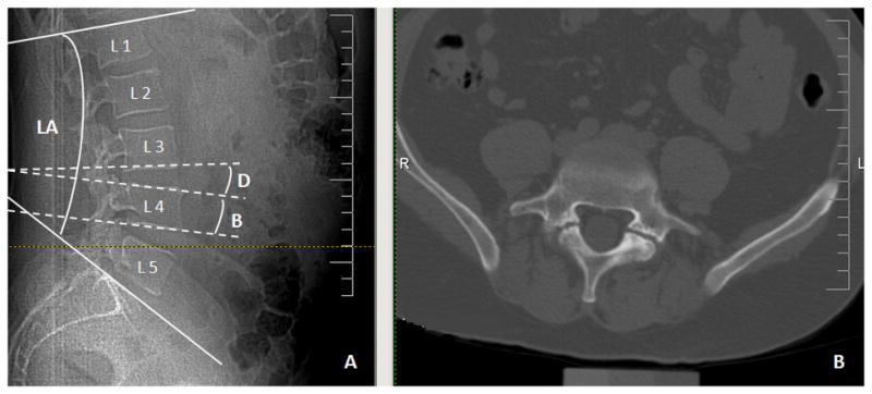
A. Measurements of lumbar angle (LA), vertebral body angle (B) and intervertebral disc angle (D).
B. Bilateral spondylolysis of L5 is shown on axial views image.

Example of evaluated CT:
A. Measurements of lumbar angle (LA), vertebral body angle (B) and intervertebral disc angle (D).
B. Bilateral spondylolysis of L5 is shown on axial views image.