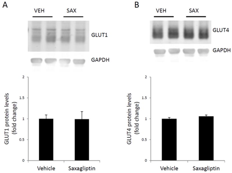
Figure 3. Western blot analysis of GLUT protein levels in left ventricular myocardium harvested from 75-day-old TG9 mice.

GLUT1 [A] and GLUT4 [B] protein levels were assessed using western blotting and normalized to the internal control, GAPDH, for loading variability. Fold change of GLUT expression in the saxagliptin-treated compared to the vehicle-treated mice is represented in the bar graphs. A representative western blot is shown. GLUT1 and GLUT4 levels are unchanged in left ventricular myocardium following sustained treatment with saxagliptin. Values are expressed as the mean ± SEM. n = 6 mice per group.
