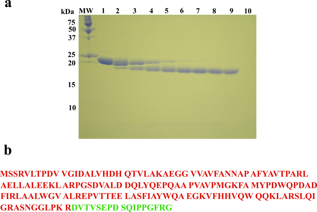
Figure 4.
a. The SDS polyacrylamide gel (15%) of the DnaT protein subjected to the time-dependent trypsin digestion (buffer C, pH 7.0, 20°C) and stained with Coomasie Brilliant Blue (Materials and Methods). Lane MW contains the molecular markers. Lane 1 contains DnaT alone. Subsequent lanes contains DnaT-trypsin mixture (molar ratio = 140:1) at different times of the digestion reaction (min); lane 2, 5; lane 3, 10; lane 4, 20; lane 5, 30; lane 6, 45; lane 7, 60; lane 8, 90; lane 9, 120; lane 10, trypsin alone (details in text). b. Primary structure of the DnaT monomer with the N-terminal core domain and the small C-terminal region marked in different colors (15).

