
Figure 6.
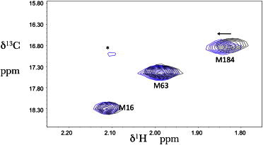
Effect of P1L on the 1H-13C HSQC spectrum of RT(I63M). 1H-13C HSQC spectra of 50 μM [13CH3-Met]66RT(I63M66) in the absence (black) and presence (blue) of 0.8 mM P1L. The resonance marked with an asterisk is unassigned but is sufficiently near the random coil values to suggest the presence of a small amount of unfolded protein. The arrow indicates the direction of the shift perturbation resulting from the P1L addition. The sample was prepared in NMR buffer (20 mM Tris-HCl-d11 in D2O, pD = 7.42, 150 mM KCl, 4 mM MgCl2 0.02% NaN3) and measured at 25°C.