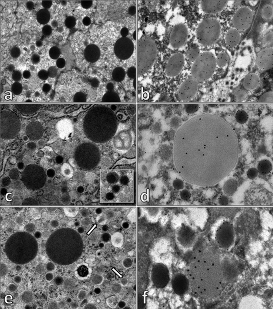
Figure 5. Electron microscopy.
The cytoplasm of acinar cells shows numerous electron dense zymogen granules (a), which are positive for trypsin (b). Some cells showed in the cytoplasm, zymogen granules and alpha-type secretory granules (c), with typical electron dense core and clear halo (inset). Double label electron microscopy immunocytochemistry (d) demonstrated that alpha-type secretory granules were glucagon-positive (glucagon immunoreactivity identified with 12 nm colloidal gold), while zymogen granules were positive for trypsin (trypsin-immunoreactivity is evidenced with 18 nm colloidal gold). Moreover, other cells presented in the cytoplasm zymogen granules were crystalline beta-type secretory granules (some indicated with arrows) (e). Double label electron microscopy immunocytochemistry (f) demonstrated that beta-type secretory granules were insulin-positive (insulin immunoreactivity identified with 12 nm colloidal gold), while zymogen granules were positive for trypsin (trypsin-immunoreactivity is evidenced with 18 nm colloidal gold). In tissues processed for electron microscopy immunocytochemistry (embedded in London White Resin, without post-fixation in 1% osmium tetroxide) zymogen granules appear clearer than that observed in tissue processed for conventional electron microscopy.

