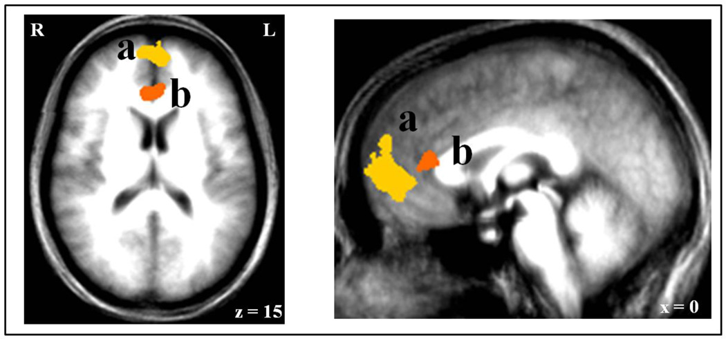
Figure 3. Regions of vmPFC identified by the analyses.
For comparison, the vmPFC showing an increased BOLD response to (a) positive relative to negative events (8, 48, −3), and the rACC showing an increased BOLD response to (b) positive events as a function of the subject’s increased probability estimates (5, 32, 12).

