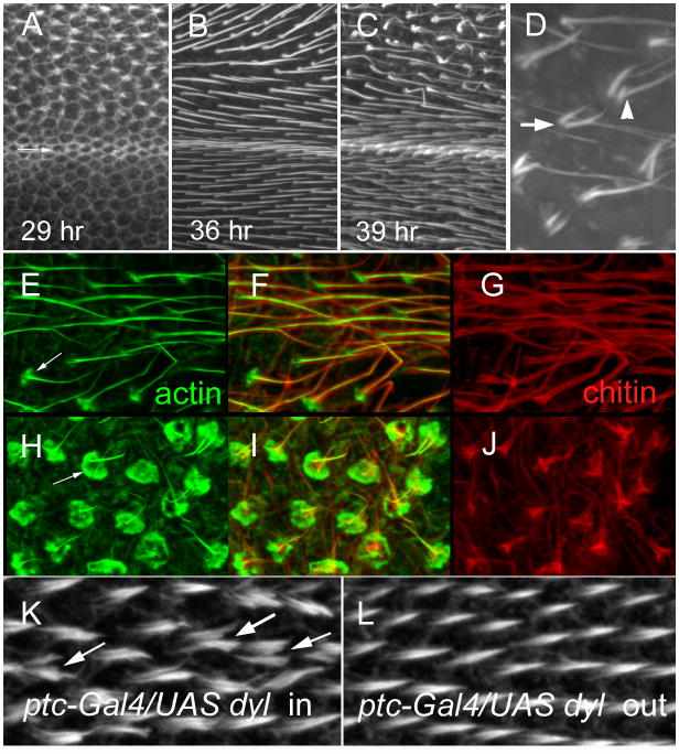
Fig. 3.
Dyl and hair morphogenesis. (A) A 29 hr ptc-Gal4 UAS-dyl RNAi pupal wing stained to show F-actin. The arrow shows the boundary between the ptc domain and the wild type wing posterior to it (below). Note the hairs forming inside the ptc domain and not outside it. (B) A 36 hr ptc-Gal4 UAS-dyl RNAi pupal wing stained for F-actin. Hairs are seen both in and outside of the ptc domain, but note the hairs inside the ptc domain appear longer, thicker and are stained more brightly. (C). A 39 hr ptc-Gal4 UAS-dyl RNAi pupal wing stained for F-actin. Note hairs inside the ptc domain are starting to appear abnormal. (D). A higher magnification view of abnormal ptc-Gal4 UAS-dyl RNAi hairs in a 39 hr wing. The arrow points to a multiple hair cell. The arrowhead points to a split hair. (E) A 43 hr ptc-Gal4 UAS-dyl RNAi pupal wing stained for F-actin. This image is for a region outside of the ptc domain. Note the F-actin (green) in the hair is central to chitin (red). Relatively weak staining of hair cups is visible (arrow). (F) A merged image of E and G. (G). The same wing region shown in E, but stained for chitin in red. (H) A 43 hr ptc-Gal4 UAS-dyl RNAi pupal wing stained for F-actin. This image is for a region inside of the ptc domain. The arrow points to the large accumulation of F-actin at the base of the hair and the abnormal structure of the hairs. (I) A merge of H and J. (J) The same wing region shown in H, but stained for chitin in red. Note the staining is far brighter in the proximal part of the hair. This is not seen in wt. (K). A 33 hr ptc-Gal4 Tub-Gal80ts UAS-dyl pupal wing inside of the ptc domain. The arrow points to a multiple/split hair cell. (L). A 33 hr ptc-Gal4 Tub-Gal80ts UAS-dyl pupal wing outside of the ptc domain. Note that the relative total hair F-actin staining is on average slightly stronger in K than L.

