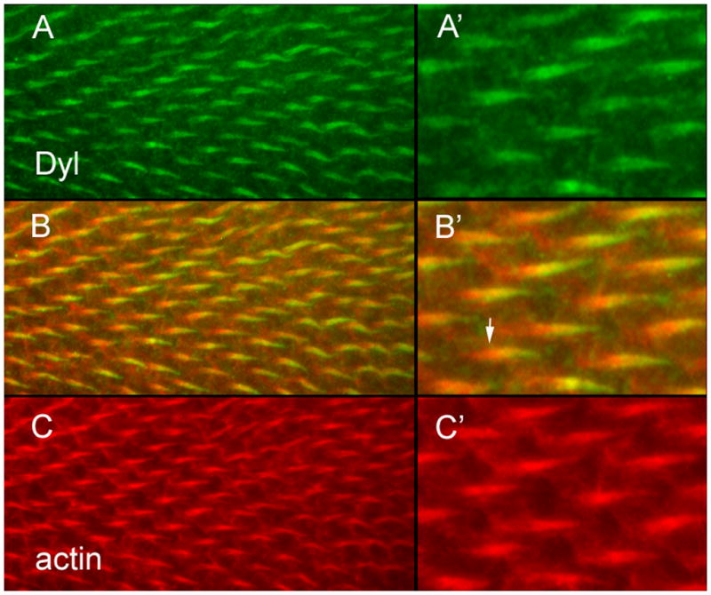
Fig. 4.

Dyl accumulates in growing hairs. (A) Dyl antibody staining shows the protein accumulates in growing hairs. (B) A merge of A and C. (C) F-actin staining. (A’) A higher magnification image of part of the field in A. (B’) A higher magnification image of part of the field in B. The arrow points to the proximal “root” of the hair that stains for actin but not the plasma membrane localized Dyl. (C’) A higher magnification image of part of the field in C.
