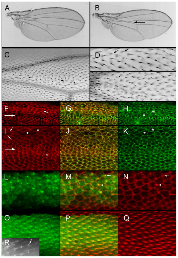
Fig. 5.
Rab11 and wing development. (A) A ptcGal4 Gal80ts/UAS-DN-Rab11 wing from a fly grown at 21°C. (B) A ptcGal4 Gal80ts/UAS-DN-Rab11 wing from a fly grown at 27.5°C. The arrow points to the reduced size of the ptc domain. (C) A micrograph of a ptcGal4 Gal80ts/UAS-DN-Rab11 wing from a fly grown at 27.5°C. Just distal to the anterior cross vein (ACV) is a group of cells showing abnormal hair polarity (arrows) and multiple hair cells (arrowheads). (D). A micrograph of a ptcGal4 Gal80ts/UAS-DN-Rab11 wing from a fly grown at 21°C until wpp and then shifted to 27.5°C. Note the presence of several thin split hairs (arrows). (E). A micrograph of a ptcGal4 Gal80ts/UAS-DN-Rab11 wing from a fly grown at 21°C until wpp and then shifted to 27.5°C. Note the lack of precise alignment of neighboring hairs. (F). A micrograph of a ptcGal4 Gal80ts/UAS-DN-Rab11 pupal wing from a fly grown at 27.5°C. The large arrow shows the boundary of the ptc domain (above inside). This image shows immunolocalization of Stan. The small arrow shows a cell with an increased level of improperly localized Stan. (G). A merge of F and H. (H). The same wing shown in F but showing F-actin staining in green. The asterisks are on abnormally shaped cells. (I). A micrograph of a ptcGal4 Gal80ts/UAS-DN-Rab11 pupal wing from a fly grown at 27.5°C. The large arrow shows the boundary of the ptc domain (above inside). This image shows immunolocalization of In. Note the zigzag accumulation pattern for In outside of the ptc domain and the aberrant cell shape and In localization inside the ptc domain. (J). A merge of I and K. (K). The same wing as in I but showing F-actin staining. The asterisks are on abnormally shaped cells. (L). A 32 hr ptc-Gal/UAS-GFP-Rab11 pupal wing. (M) A merge of L and N. The arrow points to a cell where GFP-Rab11 is showing distal accumulation prior to F-actin accumulation. This shows that GFP-Rab11 is an earlier marker of hair outgrowth than F-actin. The arrowhead points to a cell where the hair is also marked by F-actin accumulation. (O). A 34 hr ptc-Gal/UAS-GFP-Rab11 pupal wing stained for GFP. (P) A merge of O and Q. (Q). A 34 hr ptc-Gal/UAS-GFP-Rab11 pupal wing stained for F-actin. (R) A 35 hr ap-Gal/UAS-GFP-Rab11 pupal wing imaged in vivo. The arrow points to the “blob” of GFP-Rab11 at the tip of the growing hair.

